Blank Diagram Of A Long Bone - Muscular And Skeletal Systems - The structure of a long bone allows for the best visualization of all of the parts of a bone (figure 1).
Blank Diagram Of A Long Bone - Muscular And Skeletal Systems - The structure of a long bone allows for the best visualization of all of the parts of a bone (figure 1).. Labeled diagram of an osteon. Long bones function as rigid bars that move when muscles contract. Long bones include the humerus (upper arm), radius (forearm), ulna (forearm), femur (thigh), fibula (thin bone of the lower leg), tibia (shin bone) , phalanges (digital bones in the hands and feet), metacarpals (long bones within the hand), and metatarsals (long bones. You need to get 100% to score the 10 points available. Short bones provide stability and support as well as.
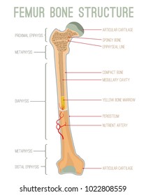
The diaphysis is the tubular shaft that runs between the proximal and distal ends of the bone. A long bone is a bone that has a shaft and 2 ends and is longer than it is wide. Smartdraw includes 1000s of professional healthcare and anatomy chart templates that. The epiphyseal line is a remnant of an area that contained hyaline cartilage that grew. Bones of the axial and appendicular skeleton.
Blank Diagram Of A Long Bone Level 3 5 Exercise And Fitness Knowledge Personal Fun Pigeon Forge from lh6.googleusercontent.com It is placed laterally to tibia and is the most slender of all the long bones. Just in case you get tired of looking at the screen we've provided images and pdf files that you can. Bones of the axial and appendicular skeleton. Short bones provide stability and support as well as. We cover the diaphysis, the epiphysis, spongy and c. The long bones have a long shaft and two bigger ends. A domino is a small tile that represents the roll of two dice. Image of a typical long bone is shown with numbers identifying the various parts, such as the epiphysis.
Related posts of diagram of of a long bone bone on side of the foot.
Long bones have a thick outside layer of compact bone and an inner medullary cavity containing bone marrow. Long bones are one of the five bone types that are classified by shape. Long blank long bone diagram bone structure diagram and metaphysisjpg from the above resolutions which is part of the human anatomydownload this image for free in hd resolution the choice download button below. Blank diagram of long bone is free hd wallpaper. Long bones function as rigid bars that move when muscles contract. The tarsus or heel bone consist of 7 bones that make up the posterior part of the foot, that is present between the tibia, fibula and metatarsals. Choose from 500 different sets of long bone diagram flashcards on quizlet. Sectional diagram of a long bone. Most, but not all, features you are required to know are shown on the following pages. Layer of bone tissue that has many small spaces and is found j…. The end of a long bone. Short bones provide stability and support as well as. It contains the bone marrow, one of the most important tissues in the vertebrate diagram of a typical long bone:
Parts of a long bone. A typical long bone shows the gross anatomical characteristics of bone. Blank diagram of a long bone. Skeletal system and long bone anatomy diagrams bundle. Long bones include the humerus (upper arm), radius (forearm), ulna (forearm), femur (thigh), fibula (thin bone of the lower leg), tibia (shin bone) , phalanges (digital bones in the hands and feet), metacarpals (long bones within the hand), and metatarsals (long bones.
Label The Parts Of A Long Bone from anatomycorner.com Bones of the axial and appendicular skeleton. The diaphysis and the epiphysis. Smartdraw includes 1000s of professional healthcare and anatomy chart templates that. The structure of a long bone allows for the best visualization of all of the parts of a bone (figure 1). Layer of bone tissue that has many small spaces and is found j…. The diaphysis and the epiphysis. A domino is a small tile that represents the roll of two dice. It is very strong to support the body's weight.
The tarsus or heel bone consist of 7 bones that make up the posterior part of the foot, that is present between the tibia, fibula and metatarsals.
Bones of the axial and appendicular skeleton. The only short bones in the human skeleton are in the carpals of the wrists and the tarsals of the ankles. It is 2 feet long and hollow, to make it lighter. Layer of bone tissue that has many small spaces and is found j…. The diaphysis is the tubular shaft that runs between the proximal and distal ends of the bone. In this video we discuss the parts of a long bone and some of the functions of each of those bone parts. A long bone has two parts: Choose from 500 different sets of long bone diagram flashcards on quizlet. Covers the surfaces of bones where they come together to form…. The end of a long bone. These include the bones of the arms and legs. There is a printable worksheet available for download here so you can take the quiz with pen and paper. Long bones are composed of both cortical and cancellous bone tissue.
The structure of a long bone allows for the best visualization of all of the parts of a bone (figure 1). Most, but not all, features you are required to know are shown on the following pages. We cover the diaphysis, the epiphysis, spongy and c. It is placed laterally to tibia and is the most slender of all the long bones. Long bones include the humerus (upper arm), radius (forearm), ulna (forearm), femur (thigh), fibula (thin bone of the lower leg), tibia (shin bone) , phalanges (digital bones in the hands and feet), metacarpals (long bones within the hand), and metatarsals (long bones.
Long Bone Anatomy Images Stock Photos Vectors Shutterstock from image.shutterstock.com Related posts of diagram of of a long bone bone on side of the foot. There is a printable worksheet available for download here so you can take the quiz with pen and paper. In long bones, as you move from the outer cortical compact bone to the inner medullary cavity, the bone transitions to spongy bone. Short bones provide stability and support as well as. Compact bone is the denser, stronger of the two types of bone tissue ( link ). Long bones function as rigid bars that move when muscles contract. This cartilage, called the epiphyseal plate, persists until the. The tarsus or heel bone consist of 7 bones that make up the posterior part of the foot, that is present between the tibia, fibula and metatarsals.
The end of a long bone.
Most, but not all, features you are required to know are shown on the following pages. Labeled diagram of an osteon. Smartdraw includes 1000s of professional healthcare and anatomy chart templates that you can modify and make your own. This cartilage, called the epiphyseal plate, persists until the. A whole skeletal muscle is considered an organ of the muscular system.each organ or muscle consists of skeletal muscle tissue, connective tissue, nerve tissue, and blood or vascular tissue. Sectional diagram of a long bone. A typical long bone shows the gross anatomical characteristics of bone. It can be found under the periosteum and in the diaphyses of long bones, where it provides support and protection. The structure of a long bone allows for the best visualization of all of the parts of a bone (figure 1). Learn long bone diagram with free interactive flashcards. This is an online quiz called label the long bone. The only short bones in the human skeleton are in the carpals of the wrists and the tarsals of the ankles. The blood vessels inside a bone.


0 Komentar